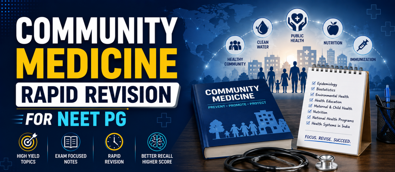
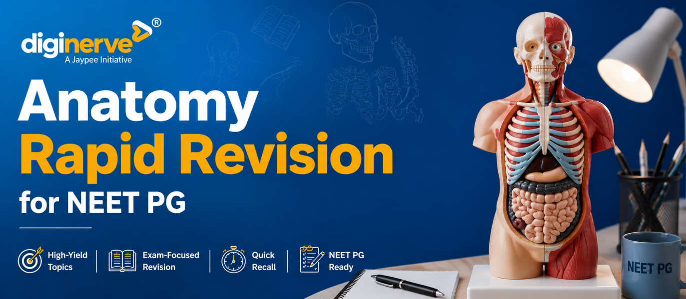
Ophthalmology Rapid Revision for NEET PG 2026: High-Yield Notes, Important Topics, PYQs & Last-Minute Tips
Preparing Ophthalmology for NEET PG 2026 requires a focused, image-based, and clinically oriented revision strategy. Ophthalmology is a scoring subject because many questions are based on common eye diseases, instruments, fundus images, visual field defects, cataract, glaucoma, retinal disorders, squint, trauma, and neuro-ophthalmology.
Ophthalmology questions in NEET PG are usually clinical, image-based, diagnosis-oriented, and instrument-based. Instead of revising lengthy theory repeatedly, aspirants should focus on high-yield topics, must-remember tables, previous year question trends, image-based questions, and rapid revision notes.

Important Topics Weightage in Ophthalmology for NEET PG
Ophthalmology in NEET PG generally includes questions from the conjunctiva, cornea, lens, glaucoma, retina, uvea, neuro-ophthalmology, squint, ocular trauma, optics, and ophthalmic instruments. Certain areas are repeatedly tested and should be prioritised during rapid revision.
| Ophthalmology Section | Importance of NEET PG |
| Cataract and Lens | Very High |
| Glaucoma | Very High |
| Retina | Very High |
| Cornea | High |
| Uvea | High |
| Neuro-Ophthalmology | High |
| Optics and Refraction | High |
| Squint and Amblyopia | Moderate to High |
| Conjunctiva | Moderate to High |
| Ocular Trauma | High |
| Ophthalmic Instruments | Very High |
| Image-Based Ophthalmology Questions | Very High |
High-Yield Ophthalmology Topics for NEET PG 2026
During the final phase of NEET PG preparation, it is important to revise the most scoring Ophthalmology topics first. These topics are commonly asked through clinical cases, fundus images, visual field charts, instruments, and surgical scenarios.
-
Cataract and Lens
Cataract is one of the most important topics in Ophthalmology for NEET PG. Focus on:
- Age-related cataract
- Congenital cataract
- Complicated cataract
- Traumatic cataract
- Diabetic cataract
- Steroid-induced cataract
- Types of cataract
- Clinical features of cataract
- Lens-induced glaucoma
- Cataract surgery
- Phacoemulsification
- Intraocular lens
- Complications of cataract surgery
- Posterior capsular opacification
- Aphakia and pseudophakia
-
Glaucoma
Glaucoma is very high-yield because questions are commonly based on clinical presentation, visual field defects, intraocular pressure, and management. Important topics include:
- Primary open-angle glaucoma
- Primary angle-closure glaucoma
- Acute congestive glaucoma
- Congenital glaucoma
- Secondary glaucoma
- Normal tension glaucoma
- Optic disc cupping
- Visual field defects
- Tonometry
- Gonioscopy
- Perimetry
- Medical management of glaucoma
- Laser iridotomy
- Trabeculectomy
- Drugs causing angle closure
-
Retina
Retina is a highly scoring and image-based section. Revise:
- Diabetic retinopathy
- Hypertensive retinopathy
- Retinal detachment
- Central retinal artery occlusion
- Central retinal vein occlusion
- Retinitis pigmentosa
- Age-related macular degeneration
- Macular hole
- Retinoblastoma
- Retinopathy of prematurity
- Fundus findings
- Fluorescein angiography
- OCT basics
- Laser photocoagulation
- Vitreous hemorrhage
-
Cornea
Corneal disorders are frequently asked about through red eye and ulcer-based clinical scenarios. Focus on:
- Corneal anatomy
- Corneal ulcer
- Bacterial keratitis
- Fungal keratitis
- Viral keratitis
- Dendritic ulcer
- Hypopyon corneal ulcer
- Keratoconus
- Corneal opacity
- Corneal dystrophies
- Corneal transplantation
- Dry eye disease
- Exposure keratitis
- Contact lens-related keratitis
-
Uvea
Uveitis is important because questions are often based on a painful red eye and systemic associations. Revise:
- Anterior uveitis
- Intermediate uveitis
- Posterior uveitis
- Panuveitis
- Keratic precipitates
- Cells and flare
- Posterior synechiae
- Hypopyon
- Sympathetic ophthalmia
- Endophthalmitis
- Systemic associations of uveitis
- Treatment of uveitis
-
Conjunctiva and Sclera
This section is commonly tested through clinical images and red eye conditions. Important topics include:
- Conjunctivitis
- Allergic conjunctivitis
- Trachoma
- Pterygium
- Pinguecula
- Subconjunctival hemorrhage
- Episcleritis
- Scleritis
- Vernal keratoconjunctivitis
- Ophthalmia neonatorum
- Dry eye
- Red eye differential diagnosis
-
Neuro-Ophthalmology
Neuro-ophthalmology is high-yield and often integrated with Medicine and Neurology. Focus on:
- Optic neuritis
- Papilledema
- Optic atrophy
- Visual pathway lesions
- Visual field defects
- Relative afferent pupillary defect
- Argyll Robertson pupil
- Marcus Gunn pupil
- Horner syndrome
- Third nerve palsy
- Fourth nerve palsy
- Sixth nerve palsy
- Internuclear ophthalmoplegia
- Pupillary light reflex pathway
-
Optics and Refraction
Optics is scoring when formulas and concepts are revised clearly. Revise:
- Myopia
- Hypermetropia
- Astigmatism
- Presbyopia
- Aphakia
- Retinoscopy
- Snellen chart
- Visual acuity
- Pinhole test
- Refractive errors
- Cylindrical lenses
- Spherical lenses
- Contact lenses
- Low vision aids
-
Squint and Amblyopia
Squint is commonly assessed through clinical examination and extraocular muscle involvement. Focus on:
- Concomitant squint
- Paralytic squint
- Esotropia
- Exotropia
- Hypertropia
- Cover test
- Hirschberg test
- Diplopia
- Amblyopia
- Extraocular muscles
- Nerve supply of extraocular muscles
- Duane retraction syndrome
- Brown syndrome
- Treatment of amblyopia
-
Ocular Trauma and Emergencies
Ocular trauma is important because questions are commonly case-based. Revise:
- Chemical injury
- Foreign body
- Blunt trauma
- Penetrating injury
- Hyphema
- Corneal abrasion
- Globe rupture
- Orbital blowout fracture
- Traumatic optic neuropathy
- Sympathetic ophthalmia
- Endophthalmitis
- Emergency management of ocular trauma
-
Ophthalmic Instruments and Procedures
Ophthalmic instruments are very important for image-based questions. Focus on:
- Direct ophthalmoscope
- Indirect ophthalmoscope
- Slit lamp
- Tonometer
- Schiotz tonometer
- Applanation tonometer
- Gonioscope
- Retinoscope
- Keratometer
- Perimeter
- A-scan
- B-scan
- OCT
- Fundus camera
- Trial frame and trial lenses
- Snellen chart
- Ishihara chart
Must-Remember Tables for Ophthalmology Rapid Revision
Tables are extremely useful for last-minute Ophthalmology revision because they help compare red eye conditions, glaucoma types, retinal vascular occlusions, refractive errors, and neuro-ophthalmic lesions quickly.
Painful Red Eye: Important Differentials
| Condition | Key Feature |
| Acute angle-closure glaucoma | Severe pain, halos, mid-dilated pupil, high IOP |
| Anterior uveitis | Pain, photophobia, small pupil, cells and flare |
| Corneal ulcer | Pain, watering, corneal opacity/ulcer |
| Scleritis | Severe deep pain, bluish-red congestion |
| Episcleritis | Mild discomfort, sectoral redness |
| Conjunctivitis | Discharge, irritation, conjunctival congestion |
Open-Angle vs Angle-Closure Glaucoma
| Feature | Open-Angle Glaucoma | Angle-Closure Glaucoma |
| Onset | Gradual | Sudden |
| Pain | Usually absent | Severe pain |
| Anterior chamber angle | Open | Closed |
| Pupil | Usually normal | Mid-dilated, fixed |
| Vision | Gradual loss | Sudden blurring with halos |
| IOP | Raised | Markedly raised |
| Treatment | Drugs, laser, surgery | Emergency IOP reduction, laser iridotomy |
Retinal Vascular Occlusions
| Condition | Classic Fundus Finding |
| Central retinal artery occlusion | Cherry-red spot |
| Central retinal vein occlusion | Blood and thunder appearance |
| Branch retinal artery occlusion | Sectoral retinal whitening |
| Branch retinal vein occlusion | Sectoral hemorrhages |
| Diabetic retinopathy | Microaneurysms, haemorrhages, neovascularisation |
| Hypertensive retinopathy | AV nicking, cotton wool spots, flame haemorrhages |
Refractive Errors
| Refractive Error | Correction |
| Myopia | Concave lens |
| Hypermetropia | Convex lens |
| Astigmatism | Cylindrical lens |
| Presbyopia | Convex lens for near vision |
| Aphakia | High plus lens/IOL/contact lens |
Visual Field Defects
| Lesion Site | Visual Field Defect |
| Optic nerve | Monocular vision loss |
| Optic chiasma | Bitemporal hemianopia |
| Optic tract | Contralateral homonymous hemianopia |
| Temporal lobe/Meyer loop | Contralateral superior quadrantanopia |
| Parietal lobe | Contralateral inferior quadrantanopia |
| Occipital cortex | Contralateral homonymous hemianopia with macular sparing |
Image-Based Questions in Ophthalmology for NEET PG
Image-based Ophthalmology questions are very common in NEET PG. Students should revise fundus images, slit-lamp findings, eye instruments, visual field charts, retinal lesions, corneal ulcers, and external eye photographs regularly.
Important image-based areas include:
- Diabetic retinopathy fundus
- Hypertensive retinopathy fundus
- Papilledema
- Optic atrophy
- Glaucomatous optic disc cupping
- Cherry-red spot in CRAO
- Blood and thunder fundus in CRVO
- Retinal detachment
- Retinoblastoma
- Age-related macular degeneration
- Corneal ulcer
- Dendritic ulcer
- Pterygium
- Trachoma
- Cataract types
- Hyphema
- Hypopyon
- Visual field defects
- Ishihara chart
- Snellen chart
- Slit lamp
- Ophthalmoscope
- Tonometer
- Retinoscope
Previous Year Questions Trend in Ophthalmology
Previous year questions show that NEET PG often tests Ophthalmology through clinical cases, fundus images, instruments, visual field charts, and emergency management. The trend is moving toward applied Ophthalmology, retinal images, glaucoma, neuro-ophthalmology, and common eye conditions.
Common PYQ trends include:
- Cataract
- Cataract surgery complications
- Primary open-angle glaucoma
- Acute angle-closure glaucoma
- Optic disc cupping
- Visual field defects
- Diabetic retinopathy
- Hypertensive retinopathy
- Retinal detachment
- CRAO and CRVO
- Retinoblastoma
- Corneal ulcer
- Dendritic ulcer
- Trachoma
- Pterygium
- Uveitis
- Papilledema
- Optic neuritis
- Third nerve palsy
- Squint
- Refractive errors
- Ocular trauma
- Ophthalmic instruments
Important MCQs in Ophthalmology
Q1. Which lens is used to correct myopia?
A. Convex lens
B. Concave lens
C. Cylindrical lens
D. Prism lens
Answer: B. Concave lens
Myopia is corrected using a concave lens, which diverges light rays and focuses the image on the retina.
Q2. A cherry-red spot on the fundus is classically seen in which condition?
A. Central retinal artery occlusion
B. Central retinal vein occlusion
C. Diabetic retinopathy
D. Hypertensive retinopathy
Answer: A. Central retinal artery occlusion
Central retinal artery occlusion classically shows a pale retina with a cherry-red spot at the macula.
Q3. Which condition presents with severe eye pain, halos around light, a mid-dilated pupil, and raised intraocular pressure?
A. Open-angle glaucoma
B. Acute angle-closure glaucoma
C. Conjunctivitis
D. Retinal detachment
Answer: B. Acute angle-closure glaucoma
Acute angle-closure glaucoma presents with severe pain, blurred vision, halos, a mid-dilated fixed pupil, and markedly raised intraocular pressure.
Q4. Dendritic corneal ulcer is commonly caused by:
A. Adenovirus
B. Herpes simplex virus
C. Staphylococcus aureus
D. Pseudomonas
Answer: B. Herpes simplex virus
Herpes simplex keratitis classically causes dendritic corneal ulcers.
Q5. Bitemporal hemianopia occurs due to a lesion at:
A. Optic nerve
B. Optic chiasma
C. Optic tract
D. Occipital cortex
Answer: B. Optic chiasma
Lesion of the optic chiasma causes bitemporal hemianopia due to involvement of crossing nasal retinal fibres.
Rapid Revision Notes for Ophthalmology
Here are some high-yield rapid revision points for NEET PG Ophthalmology:
- Myopia is corrected by a concave lens.
- Hypermetropia is corrected by a convex lens.
- Astigmatism is corrected by a cylindrical lens.
- Presbyopia is corrected with a convex lens for near vision.
- Acute angle-closure glaucoma presents with severe pain, halos, a mid-dilated pupil, and raised IOP.
- Primary open-angle glaucoma is usually painless and slowly progressive.
- Glaucoma causes optic disc cupping.
- CRAO shows a cherry-red spot.
- CRVO shows a blood and thunder appearance.
- Diabetic retinopathy shows microaneurysms, haemorrhages, hard exudates, and neovascularisation.
- Hypertensive retinopathy shows AV nicking, flame haemorrhages, cotton wool spots, and papilledema in severe cases.
- Retinal detachment presents with flashes, floaters, and curtain-like loss of vision.
- Retinoblastoma commonly presents with leukocoria.
- HSV keratitis causes a dendritic ulcer.
- Vitamin A deficiency can cause xerophthalmia and Bitot spots.
- Trachoma is caused by Chlamydia trachomatis.
- Pterygium is a triangular fibrovascular growth encroaching onto the cornea.
- Anterior uveitis presents with pain, photophobia, a small pupil, and cells and flare.
- Papilledema is optic disc swelling due to raised intracranial pressure.
- Optic neuritis causes painful vision loss and is associated with multiple sclerosis.
- Third nerve palsy causes ptosis, diplopia, and eye deviation down and out.
- Sixth nerve palsy causes lateral rectus weakness and horizontal diplopia.
- An optic chiasma lesion causes bitemporal hemianopia.
- A direct ophthalmoscope gives a magnified, erect image.
- An indirect ophthalmoscope gives an inverted image with a wider field of view.
- Tonometry is used to measure intraocular pressure.
- Gonioscopy is used to visualise the anterior chamber angle.
- OCT is useful for retina and optic nerve evaluation.
Last-Minute Tips to Revise Ophthalmology for NEET PG 2026
Ophthalmology revision should be visual, clinical, and table-based. In the last few weeks before NEET PG, avoid reading lengthy theory and focus on high-yield conditions, fundus images, instruments, PYQs, and image-based questions.
- Revise fundus images daily
The retina is one of the most image-based areas in Ophthalmology. Revise diabetic retinopathy, hypertensive retinopathy, CRAO, CRVO, papilledema, optic atrophy, and retinal detachment.
- Focus on glaucoma
Glaucoma is repeatedly asked in NEET PG. Revise open-angle glaucoma, angle-closure glaucoma, optic disc cupping, visual field defects, tonometry, and treatment.
- Make tables for red eye
Red eye questions are common. Compare conjunctivitis, keratitis, uveitis, acute glaucoma, episcleritis, and scleritis using symptoms, pupil findings, pain, and vision changes.
- Do not skip instruments
Ophthalmic instruments are frequently asked as image-based questions. Revise ophthalmoscope, slit lamp, tonometer, retinoscope, gonioscope, keratometer, and perimeter.
- Revise neuro-ophthalmology through diagrams
Visual pathway lesions, field defects, cranial nerve palsies, pupillary reflexes, and optic nerve disorders should be revised through diagrams.
- Practice clinical case-based MCQs
Ophthalmology questions are often based on symptoms like red eye, painful vision loss, sudden painless vision loss, diplopia, floaters, and leukocoria.
- Revise trauma and emergencies
Chemical injury, corneal foreign body, hyphema, acute glaucoma, CRAO, globe rupture, and endophthalmitis are important emergency topics.
- Solve PYQs thoroughly
PYQs help identify repeated Ophthalmology concepts and image patterns. After every PYQ, revise the related disease, clinical feature, investigation, and management.
Recommended Resources for Ophthalmology NEET PG Preparation
To strengthen your Ophthalmology preparation for NEET PG 2026, use a combination of structured video lectures, QBank practice, PYQ analysis, and rapid revision resources.
You can revise Ophthalmology with:
- DigiNerve NEET PG Courses
- Ophthalmology QBank
- Ophthalmology Previous Year Questions
- Ophthalmology One-Shot Revision Videos
- Subject-wise rapid revision notes
- Image-based question practice
- Related NEET PG PYQ blogs
- Previous subject revision blog
- Next subject revision blog
Frequently Asked Questions
Q1. What are the most important topics in Ophthalmology for NEET PG?
Ans – Cataract, glaucoma, retina, cornea, uveitis, neuro-ophthalmology, optics, squint, ocular trauma, and ophthalmic instruments.
Q2. How to revise Ophthalmology quickly for NEET PG?
Ans – Revise fundus images, red eye tables, glaucoma, cataract, instruments, visual field defects, PYQs, and image-based questions.
Q3. Which Ophthalmology topics are most repeated in NEET PG?
Ans – Glaucoma, cataract, diabetic retinopathy, hypertensive retinopathy, CRAO, CRVO, corneal ulcer, uveitis, visual field defects, and instruments.
Q4. Is rapid revision enough for NEET PG preparation?
Ans – Yes, for final revision, but combine it with MCQs, PYQs, image-based practice, and fundus image revision.