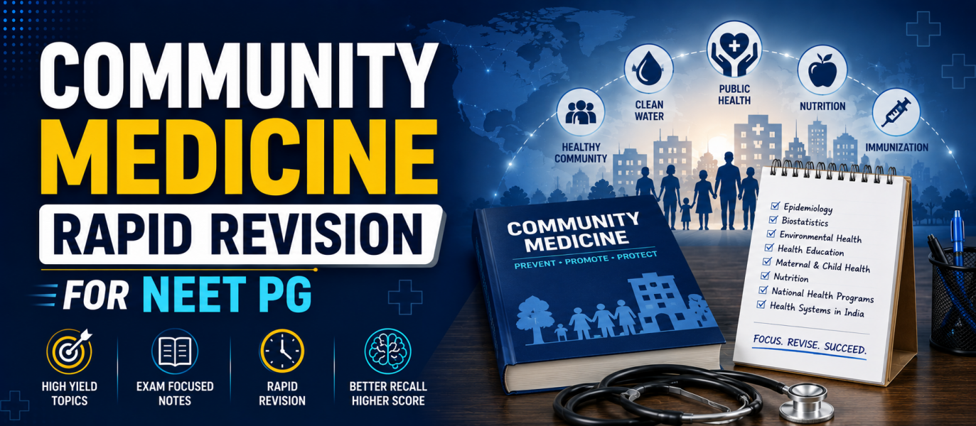
Orthopaedics Rapid Revision for NEET PG 2026
Preparing Orthopaedics for NEET PG 2026 requires a clinical, image-based, and concept-oriented revision strategy. Orthopaedics is a scoring subject because many questions focus on fractures, dislocations, nerve injuries, bone tumours, paediatric orthopaedics, arthritis, spinal disorders, trauma management, and X-ray interpretation.
Orthopaedics questions in NEET PG are usually case-based, X-ray-based, diagnosis-oriented, and management-focused. Instead of repeatedly reading lengthy theory, aspirants should focus on high-yield topics, must-remember tables, common orthopaedic images, PYQs, emergency management, and rapid revision notes.

Important Topics Weightage in Orthopaedics for NEET PG
Orthopaedics in NEET PG generally includes questions from trauma, fractures, dislocations, pediatric orthopaedics, bone tumours, infections, arthritis, spine disorders, hand injuries, nerve injuries, and orthopaedic instruments. Certain areas are repeatedly tested and should be prioritised during rapid revision.
| Orthopedics Section | Importance of NEET PG |
| Fractures and Dislocations | Very High |
| Upper Limb Injuries | Very High |
| Lower Limb Injuries | Very High |
| Pediatric Orthopedics | Very High |
| Bone Tumors | High |
| Bone and Joint Infections | High |
| Arthritis and Joint Disorders | High |
| Spine Disorders | High |
| Nerve Injuries | Very High |
| Sports Injuries | Moderate to High |
| Orthopaedic Instruments and Implants | High |
| X-ray/Image-Based Orthopaedics Questions | Very High |
High-Yield Orthopaedics Topics for NEET PG 2026
During the final phase of NEET PG preparation, it is important to revise the most scoring Orthopaedics topics first. These topics are commonly asked through clinical cases, X-rays, image-based questions, trauma scenarios, and management-based MCQs.
-
Fractures and Dislocations
Fractures and dislocations are among the most important topics in Orthopedics for NEET PG. Focus on:
- Classification of fractures
- Open and closed fractures
- Complete and incomplete fractures
- Greenstick fracture
- Stress fracture
- Pathological fracture
- Fracture healing
- Delayed union
- Non-union
- Malunion
- Complications of fractures
- Compartment syndrome
- Fat embolism syndrome
- Principles of fracture management
- Reduction and immobilization
- Internal and external fixation
- Common dislocations
- Shoulder dislocation
- Hip dislocation
- Elbow dislocation
-
Upper Limb Injuries
Upper limb injuries are high-yield because questions are often based on fracture site, nerve injury, deformity, and X-ray findings. Revise:
- Clavicle fracture
- Shoulder dislocation
- Fracture of the surgical neck of the humerus
- Fracture of the shaft of the humerus
- Supracondylar fracture of the humerus
- Lateral condyle fracture
- Medial epicondyle fracture
- Monteggia fracture-dislocation
- Galeazzi fracture-dislocation
- Colles fracture
- Smith fracture
- Scaphoid fracture
- Bennett fracture
- Mallet finger
- Jersey finger
- Dupuytren contracture
- Carpal tunnel syndrome
-
Lower Limb Injuries
Lower limb injuries are frequently asked through trauma cases and X-rays. Important topics include:
- Neck of femur fracture
- Intertrochanteric fracture
- Shaft femur fracture
- Hip dislocation
- Knee dislocation
- Patella fracture
- Tibial plateau fracture
- Shaft tibia fracture
- Ankle fractures
- Calcaneum fracture
- Metatarsal fractures
- Lisfranc injury
- ACL injury
- PCL injury
- Meniscal injury
- Achilles tendon rupture
- Foot drop
- Compartment syndrome of the leg
-
Pediatric Orthopedics
Pediatric Orthopaedics is very high-yield because questions are often based on age, deformity, X-ray findings, and congenital conditions. Focus on:
- Developmental dysplasia of the hip
- Congenital talipes equinovarus
- Perthes disease
- Slipped capital femoral epiphysis
- Supracondylar fracture in children
- Greenstick fracture
- Torus fracture
- Physeal injuries
- Salter-Harris classification
- Osteogenesis imperfecta
- Rickets
- Scurvy
- Scoliosis
- Genu varum
- Genu valgum
- Limb length discrepancy
-
Bone Tumors
Bone tumours are high-yield and commonly tested through age, site, X-ray appearance, and histology. Revise:
- Osteosarcoma
- Ewing sarcoma
- Chondrosarcoma
- Giant cell tumor
- Osteochondroma
- Osteoid osteoma
- Enchondroma
- Multiple myeloma
- Metastatic bone disease
- Aneurysmal bone cyst
- Simple bone cyst
- Chondroblastoma
- Bone tumor X-ray signs
- Codman triangle
- Sunburst appearance
- Onion-skin appearance
- Soap-bubble appearance
-
Bone and Joint Infections
Infections are commonly diagnosed through clinical signs, X-rays, and organism associations. Focus on:
- Acute osteomyelitis
- Chronic osteomyelitis
- Brodie abscess
- Septic arthritis
- Tuberculosis of bone
- Tuberculosis of the spine
- Pott spine
- Cold abscess
- Discitis
- Prosthetic joint infection
- Organisms causing bone infections
- Radiological findings in osteomyelitis
- Management of septic arthritis
-
Arthritis and Joint Disorders
Arthritis questions are often integrated with Medicine and Pathology. Revise:
- Osteoarthritis
- Rheumatoid arthritis
- Gout
- Ankylosing spondylitis
- Psoriatic arthritis
- Reactive arthritis
- Septic arthritis
- Charcot joint
- Avascular necrosis
- Frozen shoulder
- Tennis elbow
- Golfer elbow
- De Quervain tenosynovitis
- Trigger finger
- Bursitis
-
Spine Disorders
Spine disorders are high-yield because they are commonly tested through clinical features, neurological findings, and imaging. Focus on:
- Pott spine
- Scoliosis
- Kyphosis
- Lumbar disc prolapse
- Cervical spondylosis
- Lumbar canal stenosis
- Spondylolisthesis
- Ankylosing spondylitis
- Cauda equina syndrome
- Spinal cord injury
- Spinal shock
- Brown-Sequard syndrome
- Central cord syndrome
- Compression fractures
- Vertebral metastasis
-
Nerve Injuries in Orthopaedics
Nerve injuries are very important because they are repeatedly tested with fractures and dislocations. Revise:
- Axillary nerve injury
- Radial nerve injury
- Median nerve injury
- Ulnar nerve injury
- Musculocutaneous nerve injury
- Femoral nerve injury
- Sciatic nerve injury
- Common peroneal nerve injury
- Tibial nerve injury
- Erb palsy
- Klumpke palsy
- Wrist drop
- Claw hand
- Ape thumb deformity
- Foot drop
-
Orthopaedic Instruments, Implants and Procedures
Orthopaedic instruments and implants are important for image-based questions. Focus on:
- Plaster of Paris cast
- Thomas splint
- Bohler-Braun splint
- Steinmann pin
- Kirschner wire
- Intramedullary nail
- Dynamic hip screw
- Dynamic condylar screw
- External fixator
- Ilizarov fixator
- Plates and screws
- Hip prosthesis
- Knee prosthesis
- Arthroscope
- Bone grafting
- Traction methods
- Skin traction
- Skeletal traction
Must-Remember Tables for NEET PG 2026 Orthopaedics Rapid Revision
Tables are extremely useful for last-minute Orthopaedics revision because they help compare fractures, nerve injuries, tumours, pediatric conditions, and X-ray signs quickly.
Common Fractures and Associated Nerve Injuries
| Fracture/Injury | Common Nerve Injured |
| Surgical neck of the humerus fracture | Axillary nerve |
| Shaft of humerus fracture | Radial nerve |
| Supracondylar fracture of the humerus | Median nerve/anterior interosseous nerve |
| Medial epicondyle fracture | Ulnar nerve |
| Posterior hip dislocation | Sciatic nerve |
| Neck of fibula fracture | Common peroneal nerve |
| Elbow dislocation | Median/ulnar nerve |
| Colles fracture | The median nerve may be affected |
Important Fracture Eponyms
| Fracture | Key Feature |
| Colles fracture | Distal radioulnar joint with dinner fork deformity |
| Smith fracture | Reverse Colles fracture |
| Bennett fracture | Fracture-dislocation at the base of the first metacarpal |
| Monteggia fracture | Proximal ulna fracture with radial head dislocation |
| Galeazzi fracture | Distal radius fracture with distal radioulnar joint dislocation |
| Pott fracture | Bimalleolar ankle fracture |
| Jefferson fracture | Burst fracture of the atlas |
| Hangman fracture | Fracture of the pedicles of the axis |
Bone Tumours: Age, Site and X-ray Clue
| Tumor | Common Athe ge/Site | X-ray/Feature |
| Osteosarcoma | Adolescents, metaphysis Tumours of the knee | Sunburst appearance, Codman triangle |
| Ewing sarcoma | Children, diaphysis | Onion-skin appearance |
| Giant cethe tumour | Young adults, epiphysis | Soap-bubble appearance |
| Osteochondroma | Metaphysis | Bony outgrowth with cartilage cap |
| Osteoid osteoma | Young patients, cortex | Night pain relieved by NSAIDs |
| Chondrosarcoma | Adults, pelvis/shoulder | Cartilaginous tumor |
| Multiple myeloma | Elderly, axial skeleton | Punched-out lesions |
Pediatric Orthopaedic Conditions
| Condition | Key Feature |
| DDH | Positive Barlow/Ortolani test |
| CTEV | Clubfoot deformity |
| PerOrthopaedicse | Avascular necrosis of the femoral head in children |
| SCFE | Obese adolescent with hip/knee pain |
| Rickets | Bow legs, widened wrists, rachitic rosary |
| Osteogenesis imperfecta | Recurrent fractures, blue sclera |
| Salter-Harris injury | Physeal injury in children |
Arthritis and Joint Disorders
| Condition | Key Feature |
| Osteoarthritis | Joint pain worse with use, osteophytes |
| Rheumatoid arthritis | Symmetrical small joint arthritis |
| Gout | Monosodium urate crystals, podagra |
| Septic arthritis | Acute painful swollen joint with fever |
| Ankylosing spondylitis | Sacroiliitis, bamboo spine |
| Charcot joint | Neuropathic joint destruction |
| Frozen shoulder | Painful restriction of shoulder movement |
Image-Based Questions in Orthopaedics for NEET PG 2026
Image-based Orthopaedics questions are very common in NEET PG. Students should revise X-rays, clinical deformities, instruments, implants, bone tumour images, and paediatric orthopaedic images regularly.
Important image-based areas include:
- Colles fracture X-ray
- Smith fracture X-ray
- Supracondylar fracture X-ray
- Shoulder dislocation X-ray
- Hip dislocation X-ray
- Neck of femur fracture X-ray
- Intertrochanteric fracture X-ray
- Tibia fracture X-ray
- Ankle fracture X-ray
- Monteggia fracture-dislocation
- Galeazzi fracture-dislocation
- Scaphoid fracture
- Osteosarcoma X-ray
- Ewing sarcoma X-ray
- Giant cell tumor X-ray
- Multiple myeloma X-ray
- Pott spine imaging
- Osteomyelitis X-ray
- DDH imaging
- Clubfoot image
- Rickets X-ray
- Scoliosis X-ray
- Orthopedic implants
- Traction devices
- POP cast images
Previous Year Questions Trend Orthopaedics
Previous year questions show that NEET PG often tests Orthopaedics through trauma cases, X-ray interpretation, pediatric deformities, nerve injuries, bone tumours, and management decisions. The trend is moving toward applied Orthopaedics and image-based diagnosis.
Common PYQ trends include:
- Fracture healing
- Compartment syndrome
- Fat embolism
- Colles fracture
- Supracondylar fracture
- Shoulder dislocation
- Hip dislocation
- Neck of femur fracture
- Monteggia and Galeazzi fracture-dislocation
- Nerve injuries with fractures
- DDH
- CTEV
- Perthes disease
- SCFE
- Osteosarcoma
- Ewing sarcoma
- Giant cell tumor
- Osteomyelitis
- Pott spine
- Osteoarthritis
- Rheumatoid arthritis
- Lumbar disc prolapse
- Orthopaedic implants and splints
Important MCQs in Orthopaedics For NEET PG 2026
Q1. Which nerve is commonly injured in a fracture of the shaft of the humerus?
A. Axillary nerve
B. Radial nerve
C. Median nerve
D. Ulnar nerve
Answer: B. Radial nerve
The radial nerve is commonly injured in a fracture of the shaft of the humerus because it runs in the radial groove of the humerus.
Q2. Dinner fork deformity is classiseethe n in whi’ shaft ch fracture.
A. Smith fracture
B. Colles fracture
C. Scaphoid fracture
D. Bennett fracture
Answer: B. Colles fracture
Colles fracture is a distal radius fracture with dorsal displacement, producing dinner fork deformity.
Q3. Onion-skin appearance on X-ray is associated with which bone tumour?
A. Osteosarcoma
B. Giant cell tumor
C. Ewing sarcoma
D. Osteochondroma
Answer: C. Ewing sarcoma
Ewing sarcoma classically shows an onion-skin periosteal reaction on X-ray.
Q4. Positive Barlow and Ortolani tests are associated with:
A. Clan ubfoot
B. Developmental dysplasia of the hip
C. Perthes disease
D. Slipped capital femoral epiphysis
Answer: B. Developmental dysplasia of the hipThe
Barthé and Ortolani tests are used for clinical screening of developmental dysplasia of the hip in infants.
Q5. Foot drop is commonly caused by injury to which nerve?
A. Femoral nerve
B. Tibial nerve
C. Common peroneal nerve
D. Obturator nerve
Answer: C. Common peroneal nerve
Common peroneal nerve injury can cause foot drop due to weakness of ankle dorsiflexion.
Rapid Revision Notes for NEET PG Orthopaedics
Here are some high-yield rapid revision points for NEET PG Orthopaedics:
- Fracture of the shaft of the humerus is only associated with radial nerve injury.
- A surgical neck of the humerus fracture may injure the axillary nerve.
- A supracondylar fracture of the humerus may injure the median nerve or anterior interosseous nerve.
- The neck of the fibula supracondylar may injure the common peroneal nerve.
- Colles fracture causes dinner fork deformity.
- Smith fracture is a reverse Colles fracture.
- Monteggia fracture is a proximal ulna fracture with radial head dislocation.
- Galeazzi fracture is a distal radius fracture with distal radioulnar joint dislocation.
- A scaphoid fracture may cause avascular necrosis of the proximal pole.
- Posterior hip dislocation may injure the sciatic nerve.
- Compartment syndrome presents with severe pain, pain on passive stretch, paresthesia, pallor, pulselessness, and paralysis.
- Fat embolism may occur after long bone fractures.
- Osteosarcoma shows a sunburst appearance and a Codman triangle.
- Ewing sarcoma shows an onion-skin appearance.
- Giant cell tumour shows a papule-bubble appearance.
- Osteoid osteoma causes night pain relieved by NSAIDs.
- Multiple myeloma shows punched-out bone lesions.
- DDH is screened using the Barlow and Ortolani tests.
- CTEV is also known as clubfoot.
- Perthes disease is avascular necrosis of the femoral head in children.
- SCFE is common in obese adolescents with hip or knee pain.
- Pott’s spine is tuberculosis of the spine.
- Osteoarthritis shows joint space narrowing, osteophytes, subchondral sclerosis, and cysts.
- Ankylosing spondylitis may show a bamboo spine.
- Rheumatoid arthritis commonly affects small joints symmetrically.
- Common peroneal nerve injury causes foot drop.
- Radial nerve injury causes wrist drop.
- Median nerve injury can cause ape thumb deformity.
- Ulnar nerve injury can cause claw hand.
- Thomas splint is used for femur fractures.
- The dynamic hip screw is commonly used for an intertrochanteric fracture.
Last-Minute Tips to Revise OrthopedThe dynamic NEET PG 2026
Orthopedics revisian on should be image-based, trauma-focused, and table-oriented. In the last few weeks, Orthopaedics avoid reading lengthy theory and focus on high-yield fractures, X-rays, nerve injuries, bone tumours, pediatric conditions, and PYQs.
- Revise fracture X-rays daily
Orthopaedics is highly image-based. Radiographs of Colles fracture, supracondylar fracture, neck femur fracture, hip dislocation, Monteggia fracture, Galeazzi fracture, and ankle fractures.
- Make a nerve injury table
Nerve injury associations are repeatedly asked. Revise the fracture site, nerve injured, deformity, and motor/sensory loss together.
- Focus on pediatric orthopedithe cs
DDH, CTEV, Perthes disease, SCFE, rickets, osteogenesis imperfecta, and Salter-Harris injuries are common NEET PG topics.
- Revise bone tumours using age-site-X-ray pattern
Bone tumors become easier when revised through classic triads: age group, bone site, and X-ray appearance.
- Do tumour emergencies
Compartment syndrome, open fractures, septic arthritis, cauda equina syndrome, fat embolism, and dislocations require urgent diagnosis and management.
- Practice image-based questions
Revise clinical deformities, orthopaedic instruments, implants, traction devices, POP casts, and X-ray findings.
- Use PYQs to identify orthopaedics
PYQs help recognise commonly repeated fracture patterns, pediatric disorders, and tumor X-ray signs.
- Attempt the MCQs to recognise
Orthopaedics is retained better through active recall. Solve MCtumourily and revise incorrect answers with Orthopaedics-rays and tables.
Frequently Asked Questions
Q1. What are the most important topics in Orthopaedics for NEET PG?
Ans – Fractures, dislocations, nerve injuries, pediatric orthopaedics, bone tumours, Orthopaedics, Pott spine, arthritis, spine disorders, and orthopedic instruments.
Q2. How to revitumourshopedics quickly for NEET PG?
Ans- Revise X-rays, fracture tables, orthopaedic injury associations, bone tumour signs, pediatric orthopaedics, PYQs, and image-based questions.
Q3. Which Orthopaedics topics are most relevant in NEET PG?
Ans – Orthopaedics, supracondylar fracture, humerus shaft fracture, dislocation, DDH, CTEV, Perthes disease, SCFE, osteosarcoma, Ewing sarcoma, and nerve injuries.
Q4. Is rapid revision enough for NEET PG preparation?
Ans – Yes, for final revision, but combine it with MCQs, PYQs, X-ray interpretation, and image-based practice.