Abrasion: Types, Causes, Symptoms, Diagnosis and Treatment
An abrasion is a superficial traumatic injury produced when friction, scraping, compression or tangential blunt force removes the epidermis and, in more severe cases, exposes part of the underlying dermis. Abrasions are among the most frequently encountered injuries in emergency medicine, general practice, sports medicine, forensic medicine and trauma care.
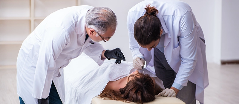
Although many abrasions are clinically minor, their appearance may provide important information regarding the mechanism, direction and nature of trauma. In forensic medicine, the site, shape and pattern of an abrasion may help reconstruct an injury and may occasionally reproduce the surface characteristics of the causative object.
From a clinical perspective, the major concerns in abrasion management are adequate wound cleansing, removal of embedded foreign material, prevention of infection, evaluation of tetanus immunisation status and recognition of associated injuries. Abrasions involving deeper dermal tissue, large surface areas, contaminated wounds or anatomically sensitive sites require more careful medical assessment.
What Is an Abrasion?
An abrasion is an injury caused by the removal of the superficial layers of the skin through mechanical friction or scraping. The term is generally used for injuries involving the epidermis, although deeper abrasions may extend into the papillary or reticular dermis.
The clinical appearance depends on:
- The force applied
- Direction of movement
- Roughness of the causative surface
- Duration of contact
- Anatomical location
- Thickness of the epidermis
- Degree of contamination
- Presence of associated blunt or penetrating trauma
Abrasions may be nearly painless when extremely superficial, but many are painful because the epidermal barrier is disrupted and dermal sensory nerve endings become exposed. Mild capillary bleeding or serous exudation may occur when the injury reaches the dermoepidermal junction or superficial dermis.
Most epidermis-limited abrasions heal through re-epithelialisation without permanent scarring. When the injury extends into the deeper dermis, healing may involve granulation tissue formation, fibrosis and scar formation.
Relevant Anatomy of the Skin
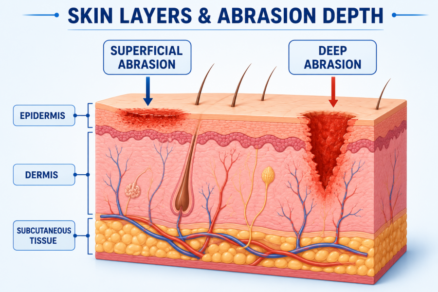
Understanding the structure of the skin helps explain the clinical behaviour of abrasions.
Epidermis
The epidermis is the outer avascular epithelial layer of the skin. It is composed primarily of keratinocytes and is organised into the following layers:
- Stratum basale
- Stratum spinosum
- Stratum granulosum
- Stratum lucidum in thick skin
- Stratum corneum
The basal layer contains proliferating keratinocytes responsible for epidermal renewal. Superficial abrasions that preserve the basal layer and skin appendages generally heal rapidly through keratinocyte migration and proliferation.
Dermis
The dermis is a vascular connective-tissue layer located beneath the epidermis. It consists of:
- Papillary dermis
- Reticular dermis
It contains collagen, elastin, blood vessels, lymphatic vessels, sensory receptors, hair follicles, sweat glands and sebaceous glands.
When an abrasion extends into the dermis, small vessels may rupture, producing pinpoint or diffuse bleeding. Damage to deeper dermal collagen increases the likelihood of fibrosis and scar formation.
Skin Appendages and Re-Epithelialisation
Hair follicles, sweat ducts and sebaceous glands contain epithelial cells that can contribute to wound re-epithelialisation. Preservation of these structures supports rapid healing. Destruction of deep dermal appendages may result in slower healing and greater scarring.
Pathophysiology of Abrasion
An abrasion is typically caused by tangential force acting parallel to the surface of the skin. This produces shearing, friction and mechanical separation of epidermal cells.
The pathological process may be described in the following sequence:
- A rough or pointed object contacts the skin.
- Friction or scraping disrupts the stratum corneum.
- Continued force removes deeper epidermal layers.
- Injury reaching the dermoepidermal junction exposes capillaries and sensory nerve endings.
- Platelets aggregate and a fibrin clot forms.
- Local inflammatory mediators initiate vasodilation and leukocyte recruitment.
- Keratinocytes migrate across the wound surface.
- The epidermal barrier is restored through proliferation and differentiation.
- Deeper injuries undergo collagen deposition and tissue remodelling.
Abrasions caused by road accidents may have particles of gravel, asphalt, soil, glass or metal embedded within the dermis. If these particles are not removed before re-epithelialisation, they may produce permanent pigmentation known as traumatic tattooing.
Types of Abrasion
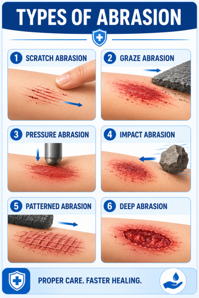
Abrasions may be classified according to their morphology, mechanism, depth and forensic characteristics.
-
Scratch Abrasion
A scratch abrasion is produced by a sharp or pointed object moving across the surface of the skin.
Common causative objects include:
- Fingernails
- Thorns
- Needles
- Pins
- Animal claws
- Sharp wires
- Rough edges
Scratch abrasions are usually linear and may be narrow at the beginning and broader at the terminal end. The accumulation of loosened epidermis at one end may sometimes suggest the direction in which the causative object moved.
Fingernail abrasions may appear as:
- Linear scratches
- Crescent-shaped marks
- Curved abrasions
- Multiple parallel abrasions
Their interpretation must be cautious because skin movement, post-injury handling and healing can alter their appearance.
-
Graze or Brush Abrasion
A graze abrasion is caused by tangential friction between the skin and a broad, rough surface. It is also called a sliding abrasion or brush abrasion.
Common examples include:
- Falling on a road
- Motorcycle or bicycle accidents
- Sliding on concrete
- Sports-field injuries
- Turf burns
- Dragging injuries
The severity depends on:
- Speed of movement
- Force of impact
- Distance travelled across the surface
- Roughness of the contact surface
- Protective clothing
- Anatomical area involved
A graze abrasion may show parallel linear marks caused by irregularities in the surface. Tags of epidermis may collect at the terminal end and occasionally indicate the direction of movement.
-
Pressure Abrasion
A pressure abrasion is produced when the skin is compressed against an underlying firm structure. The epidermis is damaged by crushing and local friction.
Examples include:
- Ligature marks
- Tight straps
- Seat-belt abrasions
- Rope compression
- Pressure from a heavy object
- Compression against bony prominences
Pressure abrasions may initially appear pale because of local vascular compression. They may later become reddish-brown as tissue dries and inflammatory changes develop.
-
Impact Abrasion
An impact abrasion occurs when a blunt object strikes the skin approximately perpendicular to its surface. The epidermis is crushed between the object and underlying tissues.
The abrasion may reproduce the shape or pattern of the causative object, particularly when the force is concentrated over a textured surface.
-
Patterned or Imprint Abrasion
A patterned abrasion carries a recognisable impression of the object responsible for the injury.
Examples include:
- Tyre-tread marks
- Radiator-grille marks
- Rope patterns
- Seat-belt marks
- Fabric impressions
- Shoe-sole patterns
- Textured tool marks
Patterned abrasions can have forensic significance because they may help correlate an injury with a suspected object. However, skin elasticity, movement and tissue distortion may affect the accuracy of the reproduced pattern.
-
Superficial Abrasion
A superficial abrasion involves only the upper epidermal layers.
Clinical characteristics include:
- Red or pink appearance
- Minimal bleeding
- Mild burning sensation
- Limited serous discharge
- Rapid healing
- Low risk of scarring
These abrasions usually heal through epidermal regeneration.
-
Deep Abrasion
A deep abrasion extends into the dermis.
Clinical characteristics include:
- More severe pain
- Capillary bleeding
- Greater tissue exudation
- Embedded foreign bodies
- Delayed epithelialisation
- Risk of infection
- Pigmentary changes
- Scar formation
Deep road-rash injuries may occasionally resemble partial-thickness burns.
Causes of Abrasion
Falls
Falls on concrete, gravel, tiled floors or uneven surfaces commonly cause abrasions of the knees, elbows, palms and face.
Road-Traffic Accidents
Motorcycle, bicycle and pedestrian accidents may produce extensive abrasions. These injuries are frequently contaminated with road debris and may coexist with fractures, head injuries or internal trauma.
Sports Injuries
Contact sports and activities involving rapid movement may cause abrasions through:
- Player-to-player contact
- Sliding on artificial turf
- Falls on hard surfaces
- Contact with sports equipment
Occupational Trauma
People working in construction, factories, agriculture or mechanical industries may be exposed to rough surfaces, rotating machinery, wires and sharp equipment.
Animal-Related Injuries
Animal claws may produce scratch abrasions. These wounds may also introduce bacteria and must be assessed for possible bite-related infection or rabies exposure, depending on the nature of the contact.
Repetitive Friction
Repeated rubbing by clothing, footwear, or equipment may produce friction abrasions. Examples include:
- Poorly fitting shoes
- Sports gear
- Repeated contact with straps
- Prolonged running or cycling
- Medical-device friction
Physical Assault
Fingernail scratches, dragging injuries, ligature abrasions and patterned abrasions may be seen in physical assault. Their location and morphology require careful documentation.
Clinical Features and Symptoms of Abrasion
The presentation depends on the depth, area, mechanism and degree of contamination.
Local Symptoms
Common local symptoms include:
- Pain
- Burning or stinging
- Tenderness
- Redness
- Mild swelling
- Raw skin surface
- Serous oozing
- Pinpoint bleeding
- Foreign-body sensation
- Crusting or scab formation
Pain may be disproportionately severe in a superficial abrasion because of exposure of sensory nerve endings.
Signs Suggesting Deeper Injury
Features that may indicate deeper tissue involvement include:
- Persistent bleeding
- Visible subcutaneous tissue
- Loss of sensation
- Reduced movement
- Severe swelling
- Deformity
- Exposed tendon or bone
- Wound contamination extending below the dermis
Signs of Infection
Clinical indicators of wound infection include:
- Increasing pain after initial improvement
- Expanding erythema
- Local warmth
- Progressive swelling
- Purulent discharge
- Malodour
- Delayed healing
- Fever
- Lymphangitic streaking
- Regional lymphadenopathy
Contaminated deep scrapes and wounds containing foreign material carry a greater risk of infection.
Colour Changes and Healing Appearance
The surface of a fresh abrasion may initially appear bright red because of exposed capillaries. It subsequently becomes covered by dried blood, fibrin and inflammatory exudate.
A general sequence may include:
- Fresh injury: bright red and moist
- Early drying: reddish-brown surface
- Crust formation: dark brown scab
- Epithelialisation: scab begins to separate
- Healed abrasion: pink or hypopigmented area
- Late healing: gradual restoration of normal pigmentation
The appearance and healing rate are influenced by wound depth, infection, anatomical location, age, circulation and comorbidities. Visual estimation should not be used alone to determine the precise age of an injury.
Diagnosis of Abrasion
Diagnosis is usually clinical and is based on history and physical examination. Abrasions do not routinely require laboratory testing or imaging unless there is concern about contamination, infection, foreign material or associated trauma.
Clinical History
Important questions include:
- When did the injury occur?
- What caused it?
- Was the skin dragged across a surface?
- Was the wound contaminated with soil, road material or animal saliva?
- Was there loss of consciousness?
- Is movement restricted?
- Is there numbness?
- Has first aid already been performed?
- What is the patient’s tetanus immunisation status?
- Does the patient have diabetes, vascular disease or immunosuppression?
Physical Examination
The clinician should assess:
- Anatomical location
- Length and surface area
- Depth
- Shape and pattern
- Directional features
- Presence of debris
- Bleeding
- Neurovascular status
- Tendon function
- Joint involvement
- Associated lacerations
- Bruising or swelling
- Signs of fracture
- Infection
Wound Exploration
Adequate illumination and pain control are important. The wound should be gently irrigated and inspected for:
- Gravel
- Glass
- Metal fragments
- Wood
- Asphalt
- Devitalised tissue
Imaging
Imaging is not routinely required for a simple abrasion. It may be indicated when there is suspicion of:
- Fracture
- Joint penetration
- Radiopaque foreign body
- Deep glass or metal fragment
- Head injury
- Internal trauma
Plain radiography may detect metal, glass and some gravel. Ultrasound may be useful for selected radiolucent foreign bodies.
Laboratory Investigations
Laboratory tests may be considered when there is:
- Extensive trauma
- Systemic infection
- Uncontrolled bleeding
- Diabetes with poor wound healing
- Suspected anaemia
- Immunocompromised status
- Severe cellulitis or sepsis
Wound cultures are generally not required for uncomplicated abrasions. They may be obtained from clinically infected wounds, recurrent infections or wounds not responding to empirical therapy.
Differential Diagnosis
Conditions that may resemble or accompany an abrasion include:
Laceration
A laceration is a tear in the skin caused by blunt or sharp trauma. Unlike a typical abrasion, it involves separation of tissue edges and may extend through the full thickness of the skin.
Incised Wound
An incised wound is produced by a sharp-edged object and generally has clean, well-defined margins.
Friction Burn
A friction burn combines mechanical abrasion with heat-induced tissue injury. It may involve deeper skin layers and sometimes requires burn-style wound management.
Contact Dermatitis
Irritant or allergic dermatitis may cause erythema, scaling and superficial erosions but generally lacks a clear traumatic mechanism.
Excoriation
Excoriations are self-induced or scratching-related erosions and may occur in pruritic skin disorders.
Pressure Injury
Pressure injuries arise from prolonged pressure and tissue ischaemia rather than brief mechanical scraping.
Erosion
An erosion is a superficial loss of epidermis that may result from infection, inflammatory dermatoses or ruptured vesicles rather than external trauma.
Wound-Healing Process in Abrasion
Abrasion healing progresses through overlapping biological phases.
-
Haemostasis
Immediately after injury:
- Small vessels constrict
- Platelets adhere to exposed tissue
- Platelet aggregation occurs
- The coagulation cascade produces fibrin
- A provisional clot seals the wound
-
Inflammatory Phase
Neutrophils migrate into the wound and remove microorganisms and debris. Macrophages subsequently coordinate repair by releasing growth factors and clearing damaged tissue.
Clinical findings during normal inflammation may include mild redness, warmth and swelling. Progressive or spreading inflammation should raise concern for infection.
-
Proliferative Phase
The proliferative phase involves:
- Keratinocyte migration
- Re-epithelialisation
- Fibroblast activity
- Collagen synthesis
- Angiogenesis
- Granulation tissue formation in deeper injuries
Superficial abrasions rely mainly on keratinocyte migration from the wound edges and preserved adnexal structures.
-
Remodelling Phase
Collagen fibres are reorganised, and tensile strength gradually increases. Deep abrasions may undergo prolonged remodelling and can leave scars or altered pigmentation.
Treatment of Abrasion
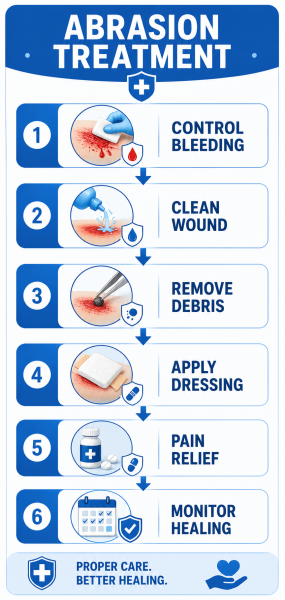
The goals of treatment are to:
- Control bleeding
- Remove contamination
- Reduce bacterial burden
- Preserve viable tissue
- Maintain a moist healing environment
- Prevent infection
- Relieve pain
- Prevent traumatic tattooing
- Assess tetanus risk
- Identify associated injuries
Initial Stabilisation
In major trauma, airway, breathing and circulation take priority over local wound care. Extensive abrasions may coexist with fractures, head injury or internal bleeding.
Haemostasis
Most abrasions cause minor bleeding that can be controlled with direct pressure using sterile gauze.
Persistent bleeding may require:
- Prolonged pressure
- Haemostatic dressing
- Evaluation for vascular injury
- Assessment for anticoagulant use or bleeding disorders
Analgesia
Wound cleansing can be painful. Pain control may involve:
- Oral analgesics
- Topical anaesthetic preparations
- Local anaesthetic infiltration in selected wounds
- Procedural analgesia for extensive road rash
Irrigation
Copious irrigation is one of the most important elements of treatment. Clean running water or normal saline can be used to remove:
- Dirt
- Clotted blood
- Microorganisms
- Loose foreign material
- Surface contaminants
The aim is mechanical cleansing rather than tissue toxicity. Aggressive scrubbing should be avoided unless necessary to remove firmly embedded particles.
Debridement
Debridement is the removal of foreign material, non-viable tissue and heavily contaminated debris.
It may be performed using:
- Sterile gauze
- Forceps
- Soft brushes
- Irrigation
- Surgical instruments
Road-rash wounds require meticulous removal of asphalt and gravel before epithelial closure to reduce the risk of traumatic tattooing.
Antiseptics
Antiseptic solutions may be used on the surrounding skin, but repeated application of highly cytotoxic substances directly into an open wound may damage viable cells and delay healing.
Routine wound irrigation should generally prioritise clean water or saline. Antiseptic selection should depend on wound characteristics and local clinical protocols.
Moist Wound Healing
Abrasions generally heal more effectively in a protected, moist environment than when repeatedly allowed to dry and crack.
A thin protective layer of petrolatum may:
- Reduce water loss
- Prevent adherence of the dressing
- Support epithelial migration
- Reduce repeated mechanical trauma
Dressings
The ideal dressing should:
- Maintain moisture
- Absorb excess exudate
- Protect against contamination
- Minimise pain during removal
- Prevent adherence
- Permit observation when required
Options include:
- Non-adherent gauze
- Semi-permeable film dressings
- Hydrocolloid dressings
- Foam dressings
- Silicone contact layers
Dressing selection depends on wound depth, exudate volume, location and contamination.
Antibiotics
Systemic antibiotics are not routinely indicated for a clean, uncomplicated abrasion.
They may be required when there is:
- Cellulitis
- Purulent infection
- Extensive contamination
- Deep tissue involvement
- Immunosuppression
- Diabetic wound
- Associated bite injury
- Systemic signs of infection
Topical antibiotics are not mandatory for every abrasion and may cause allergic contact dermatitis in susceptible individuals.
Tetanus Prophylaxis
Tetanus prevention depends on:
- Type of wound
- Degree of contamination
- Time since injury
- Vaccination history
- Immunocompromised status
Clinical wound care includes removal of dirt, foreign material and necrotic tissue. Tetanus vaccination and, in selected high-risk situations, tetanus immune globulin may be indicated according to immunisation history and wound classification. Antibiotics should not be administered solely to prevent tetanus.
Frequently Asked Questions
Q1. Is an abrasion an open wound?
Ans – Yes. An abrasion disrupts the epidermal barrier and is therefore considered an open wound, even when bleeding is minimal.
Q2. Why can a superficial abrasion be very painful?
Ans – The removal of the epidermis exposes or irritates sensory nerve endings located near the dermoepidermal junction.
Q3. Does an abrasion always bleed?
Ans – No. Very superficial abrasions may not bleed. Pinpoint bleeding occurs when superficial dermal capillaries are damaged.
Q4. Should an abrasion be left open?
Ans – Most abrasions benefit from protection with a suitable non-adherent dressing. A protected moist environment supports epithelial migration and reduces repeated trauma.
Q4. Do abrasions require sutures?
Ans – A simple abrasion does not have separated wound edges and therefore is not usually sutured. Associated lacerations may require closure.
Q5. Can an abrasion become infected?
Ans – Yes. Infection risk increases with contamination, delayed cleansing, embedded foreign material, deep tissue injury, diabetes and immunosuppression.
Q6. What is traumatic tattooing?
Ans – Traumatic tattooing is permanent skin pigmentation caused by foreign particles, such as asphalt or gravel, becoming trapped within the dermis.