Anatomy of Neck Triangles: Structures and Boundaries
The neck triangle anatomy is important for understanding the spatial organization of vital neurovascular, muscular and lymphatic structures. The cervical triangles are especially important for clinical examination, surgical intervention and radiological interpretation making them a high-yield topic for NEET PG anatomy and MBBS students.
The sternocleidomastoid muscle (SCM) is the principal anatomical landmark that divides the neck into two major triangles:
- Anterior triangle of the neck
- The posterior triangle of the neck
Each triangle contains distinct structures essential for maintaining head, neck and upper limb functions. Understanding their boundaries and contents is key for theoretical exams and practical applications in NEET PG preparation.
Anterior Triangle of the Neck
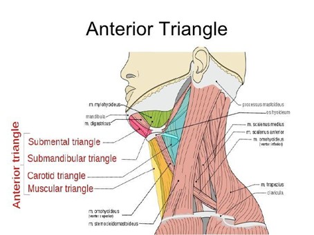
- Boundaries:
- Superior: Inferior border of the mandible
- Lateral: Anterior border of the sternocleidomastoid muscle
- Medial: Midline of the neck
- Apex: Jugular notch (at the superior border of the sternum)
- Roof: Investing layer of deep cervical fascia
- Floor: Visceral fascia overlying the pharynx, larynx and thyroid gland
- Subdivisions of the Anterior Triangle:
The anterior triangle of the neck is further divided into:
- Submental Triangle
- Submandibular (Digastric) Triangle
- Carotid Triangle
- Muscular Triangle
Each subdivision contains important vascular, lymphatic, glandular, and neural elements.
- Key Structures (The Neck Triangle Contains):
- Common carotid artery bifurcation
- Internal jugular vein
- Vagus nerve (CN X)
- The hypoglossal nerve (CN XII)
- Submandibular gland
- Thyroid and parathyroid glands
- Lymph nodes
Posterior Triangle of the Neck
- Boundaries:
- Anterior: Posterior border of the sternocleidomastoid muscle
- Posterior: Anterior border of the trapezius
- Inferior: Middle third of the clavicle
- Apex: Junction of sternocleidomastoid and trapezius near the superior nuchal line
- Roof: Investing layer of deep cervical fascia
- Floor: Prevertebral fascia covering muscles like levator scapulae and scalene group
- Subdivisions of the Posterior Triangle:
- Occipital Triangle
- Supraclavicular (Omoclavicular) Triangle
- Key Structures:
- Spinal accessory nerve (CN XI)
- Brachial plexus trunks
- Subclavian artery and vein
- Cervical lymph nodes
- Suprascapular artery
Clinical Importance in NEET PG Anatomy
Understanding the cervical triangles provides:
- Easy localisation of lymphadenopathy
- Identification of vascular injury risks (e.g., during central venous catheterisation)
- Assessment of cranial nerve damage (especially spinal accessory nerve injury in posterior triangle surgeries)
Q1. Which of the following structures lies within the carotid triangle but not in the posterior triangle of the neck?
A. Spinal accessory nerve
B. External jugular vein
C. Common carotid artery
D. Supraclavicular lymph nodes
Answer: C. Common carotid artery
Q2. During surgical dissection of the posterior triangle, a surgeon accidentally transects a nerve leading to weakness in shoulder elevation. The most likely injured structure is:
A. Greater auricular nerve
B. Spinal accessory nerve
C. Cervical branch of facial nerve
D. Ansa cervicalis
Answer: B. Spinal accessory nerve
Q3. The submandibular triangle is bounded by all of the following EXCEPT:
A. Posterior belly of digastric
B. Anterior belly of digastric
C. Stylohyoid muscle
D. Inferior border of mandible
Answer: C. Stylohyoid muscle
Q4. Which of the following triangles contains both the thyroid and parathyroid glands?
A. Carotid triangle
B. Muscular triangle
C. Submental triangle
D. Posterior triangle
Answer: B. Muscular triangle
Q5. All of the following structures form the floor of the posterior triangle EXCEPT:
A. Levator scapulae
B. Scalene medius
C. Sternocleidomastoid
D. Splenius capitis
Answer: C. Sternocleidomastoid
Q6. During central venous catheterization via the internal jugular vein, the needle inadvertently enters the carotid artery. Which triangle was most likely involved in the procedure?
A. Submandibular triangle
B. Posterior triangle
C. Carotid triangle
D. Supraclavicular triangle
Answer: C. Carotid triangle
Q7. The apex of the posterior triangle is anatomically located:
A. At the jugular notch
B. Behind the angle of the mandible
C. Where sternocleidomastoid and trapezius meet near the superior nuchal line
D. At the lateral border of the clavicle
Answer: C. Where sternocleidomastoid and trapezius meet near the superior nuchal line
Q8. Which nerve does not pass through any subdivision of the anterior triangle of the neck?
A. Hypoglossal nerve
B. Spinal accessory nerve
C. Vagus nerve
D. Ansa cervicalis
Answer: B. Spinal accessory nerve