Important Questions and Answers on Anatomy for NEET PG 2026
As the NEET PG 2026 approaches, aspirants are gearing up for India’s most important and challenging medical entrance exam. Among the numerous subjects, anatomy holds significant importance and has a considerable weight in the exam. To help you prepare efficiently, it is essential to focus on Anatomy for NEET PG, as mastering this subject can significantly enhance your chances of success. The Anatomy MCQS for NEET PG, along with Anatomy PYQS for NEET PG, play an essential role in understanding the exam pattern and the types of questions asked. Additionally, practising Anatomy questions in NEET PG will familiarise you with key concepts, making your preparation more effective.
To aid your preparation, the availability of resources such as NEET PG previous year question papers, NEET PG question banks and other resources proves invaluable. These resources not only help you gauge the level of questions asked but also provide insights into frequently asked topics. This article compiles important Anatomy questions and answers for NEET PG 2026, ensuring that you’re fully prepared to tackle this section of the exam with confidence.
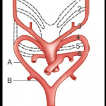
Q. 1 – Aberrant subclavian artery formed due to:
A) Persistent A
B) Persistent B
C) Persistent A and Obliterated B
D) Obliterated A and Persistent B
Ans. d. Obliterated A and persistent B
- Aberrant subclavian artery: Right fourth aortic arch and proximal portion of right dorsal aorta (‘A’) disappear, and distal portion of right dorsal aorta (‘B’) persists. In this case, the right subclavian artery is formed by the distal portion of the right dorsal aorta (‘B’) and the right seventh intersegmental artery.
- Since this abnormal artery crosses the midline behind the oesophagus and trachea, a vascular ring is formed by the right subclavian artery and the aortic arch, which may compress the two visceral tubes.
- Normally, the right subclavian artery is contributed by (proximal to distal): Right fourth arch artery, right dorsal aorta and right seventh cervical intersegmental artery.
Q. 2 – Which of the following is TRUE regarding omphalocele and gastroschisis?
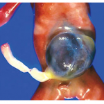
A) The herniation of abdominal contents characterises Omphalocele through a defect in the ventral abdominal wall, covered by a peritoneal membrane sac, and is often associated with multiple congenital anomalies.
B) Gastroschisis involves a midline defect, with the abdominal contents covered by a peritoneal membrane, and typically presents with an evisceration of the intestines to the right of the umbilicus.
C) Both omphalocele and gastroschisis have abdominal contents that are directly exposed to amniotic fluid.
D) Omphalocele is typically located to the right of the umbilicus and has no association with chromosomal defects.
Answer: A) The herniation of abdominal contents characterises Omphalocele through a defect in the ventral abdominal wall, covered by a peritoneal membrane sac, and is often associated with multiple congenital anomalies.
Q. 3 – A patient presented with acute abdominal pain, and on clinical suspicion patient underwent cholecystectomy. On histopathological examination, the findings are normal. The gallbladder epithelium will be:
A) Squamous
B) Simple columnar
C) Simple columnar with a brush border
D) Cuboidal with stereocilia
Ans. C. Simple columnar with brush border: Gallbladder is lined by the columnar epithelium with brush border (irregularly placed microvilli).
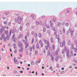
Q. 4 – Identify the organ in the following histology slide:
A) Urinary bladder
B) Gallbladder
C) Bile duct
D) Skin
Ans. A. Urinary bladder
- The slide shows transitional epithelium, which is present in the urinary tube; hence called urothelium as well.
- The most superficial cells have a thickened plasma membrane as a result of the presence of intramembranous plaques, which give an eosinophilic appearance to the luminal surface.
- Large dome-shaped (umbrella) cells that bulge into the lumen may be evident.
- Identification: At first glance, it looks like a stratified cuboidal
epithelium. Several rows of nuclei appear to be topped by a layer of dome-shaped cells which bulge into the lumen of the viscus. Cells of the basal layer are cuboidal or columnar, while the cells of the superficial layer vary in appearance depending on the degree of distension (may be squamous, if stretched).
Q. 5 – A histology slide of a gland is given diagram. Identify the type of gland:
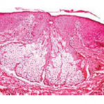
A) Apocrine
B) Merocrine
C) Holocrine
D) Endocrine
Ans. C. Holocrine
- The given slide in the figure appears to be taken from a section of skin, showing sebaceous glands.
- In holocrine glands, the secretions are produced in the cytoplasm of the cell and released by the rupture of the plasma membrane, which destroys the cell and results in the secretion of the product into the lumen.
- Examples: Sebaceous gland (skin), meibomian glands (eyelid).
Q. 6 – The following are the collagen types and their sites of location. Choose the INCORRECT pair:
A) Skin: Type I
B) Lens capsule: Type I
C) Blood vessel: Type III
D) Spleen: Type- III
E) Hyaline cartilage: Type I
Ans. B. Lens capsule: Type I e. Hyaline cartilage: type II
Generally, capsules have type-I collagen fibres, lens capsule/filtration membrane has type IV collagen fibres. Hyaline cartilage has type II collagen fibres.
Q. 7 – During the 4th week, endoderm and ectoderm approach each other in the head and neck region at:
A) Pharyngeal groove
B) Pharyngeal pouch
C) Pharyngeal membrane
D) Pharyngeal arch
Ans. C. Pharyngeal membrane
- During the 4th week, at the lateral wall of the primitive pharynx, inner endoderm (of pharyngeal pouch) and outer ectoderm (of
pharyngeal cleft) approach each other and sandwich the pharyngeal membrane between the two.
- The membrane is made up of mesenchyme (connective tissue) lined by outer ectodermal epithelium and inner endodermal epithelium.
Q. 8 – Tongue, which is NOT developed from the occipital myotome:
A) Styloglossus
B) Hyoglossus
C) Genioglossus
D) Palatoglossus
Ans. D. Palatoglossus
Tongue muscles develop from occipital myotomes except palatoglossus, which develops in pharyngeal arches.
All tongue muscles are supplied by the hypoglossal nerve except palatoglossus (supplied by the vagus accessory complex).
Q. 9 – A 59-year-old man complains of recurrent attacks in the region of the left shoulder radiating to the sternum and the pit of the stomach. The attacks of pain came at lengthy intervals until the last two days, when they became continuous. The physician diagnosed it as angina pectoris. In this case, the pain pathway from the heart is carried by:
A) Superior cervical cardiac nerve
B) Middle and inferior cervical cardiac nerve
C) Thoracic splanchnic nerve
D) Vagus
Ans. C. Thoracic splanchnic nerve
- This is a case of inferior wall MI, and the pain fibres are carried along the thoracic splanchnic nerve (greater splanchnic, T5-9), hence felt in the retrosternal and epigastric (T7) region.
- Anginal pain fibres carried by the cervical cardiac nerve may present with referred pain felt in the neck and mandible region.
Q. 10 – The following statements are true regarding the SA node except:
A) It is located at the right border of the ascending aorta
B) It contains specialised nodal cardiac muscle
C) It is supplied by the atrial branch of the right coronary artery
D) It initiates cardiac conduction
Ans. A. It is located at the right border of the ascending aorta
The SA node is located in the right atrium at the right side of the superior vena cava.
In conclusion, the key to excelling in Anatomy for NEET PG 2026 lies in consistent and targeted preparation. By incorporating Anatomy MCQS for NEET PG and solving Anatomy PYQS for NEET PG, you can ensure that you’re well-versed in the most important topics. Using the NEET PG question bank and irregularly practising NEET PG question papers will allow you to simulate the real exam environment, improving your time management skills and boosting your confidence. Stay consistent, stay focused for NEET PG 2026 preparation.